Journals > > Topics > Imaging Systems
Imaging Systems|35 Article(s)
Bionic Ultrafast Imaging for Multi-FOV and Wide Time Range
Qifan ZHU, Yi CAI, Xuanke ZENG, Hu LONG, Yongle ZHU, Liangwei ZENG, Jingzhen LI, and Xiaowei LU
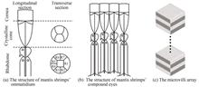
The existing ultrafast imaging systems can record dynamic events on the femtosecond time scale, but they have the problems of complex components, large systems, limited field of view and time range. The single-shot ultrafast imaging technologies by direct imaging can realize the recording of dynamic images through spatial separation of temporal images. However, their detection systems have the problem of complex structures. The ultrafast imaging system combined with algorithms simplifies the detection structure, but there are some problems, such as complex framing system, limited time range and field of view. This paper proposes a single-shot ultrafast imaging method inspired by the eyes of mantis shrimp, which can be applied to multiple fields of view and wide time range with a compact structure. In this bionic system, the structure light illumination principle is inspired by the orthogonal microvillus arrays, the ommatidium system optical path leads to the optical path of mantis shrimps' ommatidium, and the optical path of compound system is inspired by the compound eye of mantis shrimp, which expands the field of view. Single-shot bionic ultrafast imaging uses a step reflection array to generate sequential pulses of different frequencies and directions and interference fringe structured light illumination to divide the sequential images. It realizes images compressed in a single detector and reconstruction of the continuous images based on the spatial angle division multiplexing algorithm. The bionic mantis shrimp structure can realize single-shot ultrafast imaging, while the compound eye structure is composed of ommatidium systems splice, which can break through the limit size of the field of view to achieve a large field of view. The feasibility of interference fringe multiplexing is verified by static test, and Zemax establishes an optical model to realize the sequential image restoration of ommatidium and compound eye system of 8 images. The framing time is discussed and verified. FDTD simulation is used to verify the feasibility of generating sequential pulses by the step array of mirrors. The framing time can be changed by adjusting the height of adjacent steps, and achieving framing times of 78 fs and 1 ps, and the sequential pulses are not widened compared with the incident pulses. By analyzing the factors affecting the framing time of the bionic single-shot ultrafast imaging system, it can be concluded that the framing time mainly depends on the width of the sequential pulses, so its photographic frequency can reach 1013 frame/s. Besides, the time delay structure and imaging structure are independent. Dispersion element and narrowband filter array can be used instead of mirror arrays to realize time delay. As the dispersion elements can be glass columns, prism pairs, grating pairs, and optical fibers, the bionic system can expand femtosecond pulses to hundreds of femtosecond, picosecond, or nanosecond time scales. Hence the bionic ultrafast imaging system enables the recording of dynamic events on femtosecond, picosecond, or nanosecond time scales. The influence factors of the spatial resolution are then further analyzed. The optical system can achieve high spatial resolution, which can reach the pixel scale of the detector. Furthermore, it can also obtain high-quality restored images, and its intrinsic spatial resolution can theoretically reach 80.6 lp/mm. The bionic system can be used to detect various fields of view by adjusting the focal length of each lens. Hence, it can cover various fields of view. In conclusion, single-shot bionic ultrafast imaging can be applied to various sizes of the field of view in theory, and the framing rate can reach 1.2×10¹³ frame/s in simulation experiment. Single-shot bionic ultrafast imaging provides the possibility for detecting a large range of transient group events, such as light propagation in the scattering media, random motion. Its compact structure lays a foundation for miniaturization and lightweight of ultrafast imaging instruments. The existing ultrafast imaging systems can record dynamic events on the femtosecond time scale, but they have the problems of complex components, large systems, limited field of view and time range. The single-shot ultrafast imaging technologies by direct imaging can realize the recording of dynamic images through spatial separation of temporal images. However, their detection systems have the problem of complex structures. The ultrafast imaging system combined with algorithms simplifies the detection structure, but there are some problems, such as complex framing system, limited time range and field of view. This paper proposes a single-shot ultrafast imaging method inspired by the eyes of mantis shrimp, which can be applied to multiple fields of view and wide time range with a compact structure. In this bionic system, the structure light illumination principle is inspired by the orthogonal microvillus arrays, the ommatidium system optical path leads to the optical path of mantis shrimps' ommatidium, and the optical path of compound system is inspired by the compound eye of mantis shrimp, which expands the field of view. Single-shot bionic ultrafast imaging uses a step reflection array to generate sequential pulses of different frequencies and directions and interference fringe structured light illumination to divide the sequential images. It realizes images compressed in a single detector and reconstruction of the continuous images based on the spatial angle division multiplexing algorithm. The bionic mantis shrimp structure can realize single-shot ultrafast imaging, while the compound eye structure is composed of ommatidium systems splice, which can break through the limit size of the field of view to achieve a large field of view. The feasibility of interference fringe multiplexing is verified by static test, and Zemax establishes an optical model to realize the sequential image restoration of ommatidium and compound eye system of 8 images. The framing time is discussed and verified. FDTD simulation is used to verify the feasibility of generating sequential pulses by the step array of mirrors. The framing time can be changed by adjusting the height of adjacent steps, and achieving framing times of 78 fs and 1 ps, and the sequential pulses are not widened compared with the incident pulses. By analyzing the factors affecting the framing time of the bionic single-shot ultrafast imaging system, it can be concluded that the framing time mainly depends on the width of the sequential pulses, so its photographic frequency can reach 1013 frame/s. Besides, the time delay structure and imaging structure are independent. Dispersion element and narrowband filter array can be used instead of mirror arrays to realize time delay. As the dispersion elements can be glass columns, prism pairs, grating pairs, and optical fibers, the bionic system can expand femtosecond pulses to hundreds of femtosecond, picosecond, or nanosecond time scales. Hence the bionic ultrafast imaging system enables the recording of dynamic events on femtosecond, picosecond, or nanosecond time scales. The influence factors of the spatial resolution are then further analyzed. The optical system can achieve high spatial resolution, which can reach the pixel scale of the detector. Furthermore, it can also obtain high-quality restored images, and its intrinsic spatial resolution can theoretically reach 80.6 lp/mm. The bionic system can be used to detect various fields of view by adjusting the focal length of each lens. Hence, it can cover various fields of view. In conclusion, single-shot bionic ultrafast imaging can be applied to various sizes of the field of view in theory, and the framing rate can reach 1.2×10¹³ frame/s in simulation experiment. Single-shot bionic ultrafast imaging provides the possibility for detecting a large range of transient group events, such as light propagation in the scattering media, random motion. Its compact structure lays a foundation for miniaturization and lightweight of ultrafast imaging instruments.
Acta Photonica Sinica
- Publication Date: Jan. 25, 2023
- Vol. 52, Issue 1, 0111001 (2023)


Experimental Research on Calibration of the Segmented Mirror Edge Sensors Based on Point Spread Function
Bin WANG, Yichun DAI, Fangyu XU, and Zhenyu JIN
The 8-meter ring segmented primary mirror is one of the important alternatives for China Giant Solar Telescope plan. The active control technology of the segmented mirror is the key to realizing the high spatial resolution of the segmented solar telescope. In the active control of the ring segmented mirror, high precision edge detection and tip/tilt detection are important factors in determining the co-phase maintenance of the primary mirror. In the current CGST active segmented scheme, the piston error of the segmented primary mirror is detected and corrected by the electromechanical edge sensors. However, in solar observations, the daytime temperature fluctuates greatly, and the primary mirror surface is affected by solar thermal radiation to generate temperature gradients. The primary mirror temperature control is required to improve mirror seeing. The temperature of the telescope truss system will also change due to the influence of thermal radiation. Therefore, the complex observation environment of the solar telescope will cause the zero-point drift of the electromechanical edge sensor, which will gradually increase the figure error of the primary mirror and will not be able to maintain the co-phase for a long time. To solve the problem of the unstable zero-point of the electromechanical edge sensor in the solar telescope, it is necessary to find a short-period calibration method for the electromechanical edge sensor, and the calibration period is about tens of seconds to several minutes. The accuracy of edge detection is better than 5 nm, and CGST can achieve the co-phase maintenance of the primary mirror in visible or near infrared. Therefore, the calibration accuracy of the edge sensor needs to be better than 5 nm. The optical co-phase detection technology detects the figure error of the primary mirror and the phasing error, and measures the absolute position of the segments. The short-period calibration of edge sensors of segmented solar telescopes using the optical detection technology is an optional solution. In this paper, in order to verify the feasibility of short-period calibration of edge sensors with the optical detection technology, edge detection research based on point spread function is carried out. Cross-calibration experiments are carried out using actuators, edge sensors and point spread function cross-correlation detection. The detection error level of this method is evaluated, and an active control experiment based on point spread function edge detection is carried out on a two-mirror system. In the 5-hour active control experiment, the RMS of the tilt/tip change of the segmented mirror is maintained at 0.01″, the RMS of the edge height change of the segmented mirror is maintained at 6.33 nm, and the RMS of the figure error of the segmented mirror is maintained at 18.73 nm. The experimental results show that the optical edge detection can accurately reflect the change of the position state of the segments in the active control, and the edge detection accuracy is better than 5 nm. The edge detection accuracy and detection frequency based on the point spread function satisfy the short-period calibration of edge sensors. The research results provide a reference for the active maintenance of the ring segmented solar telescope in the near infrared or visible light. The 8-meter ring segmented primary mirror is one of the important alternatives for China Giant Solar Telescope plan. The active control technology of the segmented mirror is the key to realizing the high spatial resolution of the segmented solar telescope. In the active control of the ring segmented mirror, high precision edge detection and tip/tilt detection are important factors in determining the co-phase maintenance of the primary mirror. In the current CGST active segmented scheme, the piston error of the segmented primary mirror is detected and corrected by the electromechanical edge sensors. However, in solar observations, the daytime temperature fluctuates greatly, and the primary mirror surface is affected by solar thermal radiation to generate temperature gradients. The primary mirror temperature control is required to improve mirror seeing. The temperature of the telescope truss system will also change due to the influence of thermal radiation. Therefore, the complex observation environment of the solar telescope will cause the zero-point drift of the electromechanical edge sensor, which will gradually increase the figure error of the primary mirror and will not be able to maintain the co-phase for a long time. To solve the problem of the unstable zero-point of the electromechanical edge sensor in the solar telescope, it is necessary to find a short-period calibration method for the electromechanical edge sensor, and the calibration period is about tens of seconds to several minutes. The accuracy of edge detection is better than 5 nm, and CGST can achieve the co-phase maintenance of the primary mirror in visible or near infrared. Therefore, the calibration accuracy of the edge sensor needs to be better than 5 nm. The optical co-phase detection technology detects the figure error of the primary mirror and the phasing error, and measures the absolute position of the segments. The short-period calibration of edge sensors of segmented solar telescopes using the optical detection technology is an optional solution. In this paper, in order to verify the feasibility of short-period calibration of edge sensors with the optical detection technology, edge detection research based on point spread function is carried out. Cross-calibration experiments are carried out using actuators, edge sensors and point spread function cross-correlation detection. The detection error level of this method is evaluated, and an active control experiment based on point spread function edge detection is carried out on a two-mirror system. In the 5-hour active control experiment, the RMS of the tilt/tip change of the segmented mirror is maintained at 0.01″, the RMS of the edge height change of the segmented mirror is maintained at 6.33 nm, and the RMS of the figure error of the segmented mirror is maintained at 18.73 nm. The experimental results show that the optical edge detection can accurately reflect the change of the position state of the segments in the active control, and the edge detection accuracy is better than 5 nm. The edge detection accuracy and detection frequency based on the point spread function satisfy the short-period calibration of edge sensors. The research results provide a reference for the active maintenance of the ring segmented solar telescope in the near infrared or visible light.
Acta Photonica Sinica
- Publication Date: Dec. 25, 2022
- Vol. 51, Issue 12, 1211001 (2022)
Calibration and Target Position of Bionic Curved Compound Eye Composed of Multiple Cameras
Zeqiang YUAN, Yuzhang GU, Shoumeng QIU, and Xiaolin ZHANG
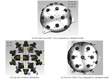
According to the characteristics of the bionic curved compound eye with many sub-eyes and multi-channel imaging, the method of multi-camera bionic curved compound eye calibration and target positioning is carried out. Combining the method of triangulation and camera coordinate system mapping under non-parallel binocular perspective, a method of bionic curved surface compound eye target positioning method is proposed. According to the requirement of compound eye positioning to have a large overlapping field of view between sub-eyes, a 17-eye bionic curved compound eye with a total field of view over 180° is designed with Unity3D simulation software, and a physical prototype is prepared. Through the ZHANG Zhengyou calibration method, the single target calibration of 17 sub-eyes in the compound eye system and the calibration between 38 pairs of adjacent sub-eyes are completed. On this basis, based on the large overlapping field of view between adjacent sub-eyes of the compound eye, the external parameter calibration method of non-adjacent sub-eyes is proposed to realize the unification of the compound eye coordinate system. Through the establishment of an experimental platform, combined with the ORB feature matching method optimized by RANSAC, a compound-eye prototype is used to perform three-dimensional positioning experiments on the unmanned aerial vehicle models at different positions, and then the error analysis of the positioning results is carried out. The experiments prove that the proposed compound eye calibration and target positioning method is applied to the prototype, which can achieve high precision and large field of view target positioning. According to the characteristics of the bionic curved compound eye with many sub-eyes and multi-channel imaging, the method of multi-camera bionic curved compound eye calibration and target positioning is carried out. Combining the method of triangulation and camera coordinate system mapping under non-parallel binocular perspective, a method of bionic curved surface compound eye target positioning method is proposed. According to the requirement of compound eye positioning to have a large overlapping field of view between sub-eyes, a 17-eye bionic curved compound eye with a total field of view over 180° is designed with Unity3D simulation software, and a physical prototype is prepared. Through the ZHANG Zhengyou calibration method, the single target calibration of 17 sub-eyes in the compound eye system and the calibration between 38 pairs of adjacent sub-eyes are completed. On this basis, based on the large overlapping field of view between adjacent sub-eyes of the compound eye, the external parameter calibration method of non-adjacent sub-eyes is proposed to realize the unification of the compound eye coordinate system. Through the establishment of an experimental platform, combined with the ORB feature matching method optimized by RANSAC, a compound-eye prototype is used to perform three-dimensional positioning experiments on the unmanned aerial vehicle models at different positions, and then the error analysis of the positioning results is carried out. The experiments prove that the proposed compound eye calibration and target positioning method is applied to the prototype, which can achieve high precision and large field of view target positioning.
Acta Photonica Sinica
- Publication Date: Sep. 25, 2021
- Vol. 50, Issue 9, 0911005 (2021)
UAV-borne Biomimetic Curved Compound-eye Imaging System for Velocity Measurement
Huangrong XU, Jinheng LIU, Yuanjie ZHANG, Dengshan WU, Hao FAN, Xiangpeng FENG, Geng ZHANG, Bingliang HU, and Weixing YU
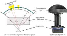
Based on the visual advantages of the biomimetic compound eye, the biomimetic curved compound-eye is applied to the unmanned aerial vehicle-borne photoelectric detection system to realize the airborne wide field-of-view and high-resolution detection of moving objects purposes. According to the characteristics of the biological compound eye, a lens array arranged on the curved surface in a hexagon is designed as compound-eye lens, combined with an optical relay subsystem and a CMOS image sensor to form a biomimetic curved compound-eye imaging and velocity measurement system. The developed biomimetic curved compound-eye imaging system has a field of view of 98°×98°, a system focal length of 5 mm, an angular resolution of 1.8 mrad and an F-number of 3.5. The size of the system is Ф123 mm ×195 mm, and the weight is 1.35 kg. According to the imaging principle of the biomimetic compound-eye and by taking advantage of the overlapping field of views between adjacent ommatidia, the velocity measurement principle of the biomimetic curved compound-eye is proposed. The velocity measurement experiment of a moving car shows that the velocity measurement method can effectively improve the test reliability and accuracy of the moving target. Based on the visual advantages of the biomimetic compound eye, the biomimetic curved compound-eye is applied to the unmanned aerial vehicle-borne photoelectric detection system to realize the airborne wide field-of-view and high-resolution detection of moving objects purposes. According to the characteristics of the biological compound eye, a lens array arranged on the curved surface in a hexagon is designed as compound-eye lens, combined with an optical relay subsystem and a CMOS image sensor to form a biomimetic curved compound-eye imaging and velocity measurement system. The developed biomimetic curved compound-eye imaging system has a field of view of 98°×98°, a system focal length of 5 mm, an angular resolution of 1.8 mrad and an F-number of 3.5. The size of the system is Ф123 mm ×195 mm, and the weight is 1.35 kg. According to the imaging principle of the biomimetic compound-eye and by taking advantage of the overlapping field of views between adjacent ommatidia, the velocity measurement principle of the biomimetic curved compound-eye is proposed. The velocity measurement experiment of a moving car shows that the velocity measurement method can effectively improve the test reliability and accuracy of the moving target.
Acta Photonica Sinica
- Publication Date: Sep. 25, 2021
- Vol. 50, Issue 9, 0911004 (2021)
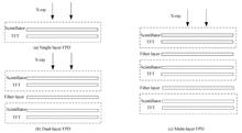
X-ray Multi-energy Imaging Method Using Multi-layer Flat Panel Detector
Zongpeng WANG, Chongzhou LAN, Minru WEN, and Chao YE
An imaging method that uses multi-layer flat panel detector to acquire X-ray multi-energy images is proposed. Single layer, multi-layer flat panel detector structure is introduced, as well as multi-layer flat panel detector detection imaging system mechanism. X-ray imaging principle is explained, dual energy imaging, subtraction principles are studied by performing single energy X-ray switching simulation with twice shots. Furthermore, different kVp, filters induced X-ray energy spectrum is analyzed, and low, medium, high energy chest phantom images are acquired by performing kVp switching experiment with several shots. Results show that multi-energy images of chest phantom exhibit difference between rib and lung region, and bone enhancement, suppression can be realized by performing dual energy subtract to multi-energy images. Similarly dual energy imaging, subtraction principles are studied by performing dual energy X-ray excited dual layer flat panel detector imaging simulation with single shot. Same kVp with different filter combination induced X-ray energy spectrum is analyzed, multi-layer flat panel detector imaging experiment with single shot is performed, and low, high energy chest phantom images are acquired by first, third layer flat panel detector. Results show that low, high energy images also exhibit difference between rib and lung region, bone enhancement, suppression can also be realized by performing dual energy subtraction. Experiment result proves that X-ray multi-energy imaging using multi-layer flat panel detector detection method is practicable. An imaging method that uses multi-layer flat panel detector to acquire X-ray multi-energy images is proposed. Single layer, multi-layer flat panel detector structure is introduced, as well as multi-layer flat panel detector detection imaging system mechanism. X-ray imaging principle is explained, dual energy imaging, subtraction principles are studied by performing single energy X-ray switching simulation with twice shots. Furthermore, different kVp, filters induced X-ray energy spectrum is analyzed, and low, medium, high energy chest phantom images are acquired by performing kVp switching experiment with several shots. Results show that multi-energy images of chest phantom exhibit difference between rib and lung region, and bone enhancement, suppression can be realized by performing dual energy subtract to multi-energy images. Similarly dual energy imaging, subtraction principles are studied by performing dual energy X-ray excited dual layer flat panel detector imaging simulation with single shot. Same kVp with different filter combination induced X-ray energy spectrum is analyzed, multi-layer flat panel detector imaging experiment with single shot is performed, and low, high energy chest phantom images are acquired by first, third layer flat panel detector. Results show that low, high energy images also exhibit difference between rib and lung region, bone enhancement, suppression can also be realized by performing dual energy subtraction. Experiment result proves that X-ray multi-energy imaging using multi-layer flat panel detector detection method is practicable.
Acta Photonica Sinica
- Publication Date: Sep. 25, 2021
- Vol. 50, Issue 9, 0911003 (2021)

Dynamic False Contour Quantification and Improvement Strategy of Digital Driven OLED
Yuan JI, Kaiwen ZHANG, Wendong CHEN, Tingzhou MU, and Shuping GONG
Aiming at dynamic false contour phenomenon may appear when digital driven OLED displays moving pictures, which affects the viewing quality of human eyes, the machanism of dynamic false contour generation is analyzed, and the visual characteristics of the human eye are considered. Just noticeable distortion integral method is proposed to quantify the dynamic false contour generated by different scanning strategies, the reliability of the evaluation method is verified through experiments. The linear pulse width modulation method and the fractal scanning method are combined, the partial fractal scanning strategy is proposed to improve the dynamic false contour phenomenon. When the data bits of the linear pulse width modulation method and the fractal scanning method are both 4 bit, the system verification of field programmable gate array is performed on a silicon-based organic light-emitting diodes microdisplay with a resolution of 1 280×1 024, and the dynamic contour phenmenon may not be perceived by the human eye. Just noticeable distortion integral method is used to evaluate the partial fractal scanning algorithm, compared with the traditional 19 subfields and fractal scanning, the average value of dynamic false contour quantization between any two gray levels is reduced by about 87.86% and 86.51%. Aiming at dynamic false contour phenomenon may appear when digital driven OLED displays moving pictures, which affects the viewing quality of human eyes, the machanism of dynamic false contour generation is analyzed, and the visual characteristics of the human eye are considered. Just noticeable distortion integral method is proposed to quantify the dynamic false contour generated by different scanning strategies, the reliability of the evaluation method is verified through experiments. The linear pulse width modulation method and the fractal scanning method are combined, the partial fractal scanning strategy is proposed to improve the dynamic false contour phenomenon. When the data bits of the linear pulse width modulation method and the fractal scanning method are both 4 bit, the system verification of field programmable gate array is performed on a silicon-based organic light-emitting diodes microdisplay with a resolution of 1 280×1 024, and the dynamic contour phenmenon may not be perceived by the human eye. Just noticeable distortion integral method is used to evaluate the partial fractal scanning algorithm, compared with the traditional 19 subfields and fractal scanning, the average value of dynamic false contour quantization between any two gray levels is reduced by about 87.86% and 86.51%.
Acta Photonica Sinica
- Publication Date: Sep. 25, 2021
- Vol. 50, Issue 9, 0911002 (2021)
Development and Application of Hyperspectral Imager Based on LVF
Qingsheng XUE, Chang LI, Tingting LI, Haoxuan BAI, Zhongtian TIAN, Bai YANG, Fupeng WANG, and Qian LI
The traditional linear variable filter hyperspectral imagers put the linear variable filter in front of the detector window. Due to the difference in the position of the linear variable filter and the detector focal plane, the spectral resolution will be reduced. In order to solve this problem, the hyperspectral imager based on linear variable filter is designed with telescope system, linear variable filter, relay system and detection system. The linear variable filter is placed on the focal plane of the telescope system, and the relay system is to image the linear variable filter on the target surface of the detector so that the linear variable filter coincides with the focal plane of the detector. For the hyperspectral imager system, the spectral range is 400~700 nm, the number of spectrum bands is 31, the spectral resolution is 10 nm, the field of view angle is ±8° and the focal length is 55 mm. After the design and construction of the sample machine, spectral calibration and application experiments is carried out with good results. Compared with traditional hyperspectral imager systems using prisms or gratings to split light, the proposed system doesn't need a collimation system, and has the advantages of small size, light weight and high luminous flux, which can provide a reference for the miniaturization of hyperspectral imagers. The traditional linear variable filter hyperspectral imagers put the linear variable filter in front of the detector window. Due to the difference in the position of the linear variable filter and the detector focal plane, the spectral resolution will be reduced. In order to solve this problem, the hyperspectral imager based on linear variable filter is designed with telescope system, linear variable filter, relay system and detection system. The linear variable filter is placed on the focal plane of the telescope system, and the relay system is to image the linear variable filter on the target surface of the detector so that the linear variable filter coincides with the focal plane of the detector. For the hyperspectral imager system, the spectral range is 400~700 nm, the number of spectrum bands is 31, the spectral resolution is 10 nm, the field of view angle is ±8° and the focal length is 55 mm. After the design and construction of the sample machine, spectral calibration and application experiments is carried out with good results. Compared with traditional hyperspectral imager systems using prisms or gratings to split light, the proposed system doesn't need a collimation system, and has the advantages of small size, light weight and high luminous flux, which can provide a reference for the miniaturization of hyperspectral imagers.
Acta Photonica Sinica
- Publication Date: Sep. 25, 2021
- Vol. 50, Issue 9, 0911001 (2021)
Research of Fiber Braking in the Single Fiber Scanning Endoscopic Imaging System
Haiyang YU, Huiying ZHANG, Simeng LIU, Jing YANG, Tianliang WANG, Bangning MAO, Yanlong MENG, Yanqing QIU, and Yi LI
An single fiber scanning endoscopic imaging system was studied in this work. A single mode fiber is driven by a piezoelectric tube with a diameter of 1 mm. The light from the fiber illuminates the target area along spiral lines, while the scattered light is collected to build an image. It is known that the precise control of the fiber movement is significantly important to the imaging quality. Researchers have found that the fiber should return back to the origin after it finished the spiral scan. Since the system can not work for imaging during the fiber return period, it is important for the scanning fiber to retreat to the origin as quick as possible. This is the key point to improve the imaging efficiency. At the same time, the residual vibrations have a great influence on the imaging quality of the subsequent scan, especially at the central area. In order to address the above problems, a theoretical mechanical model was established to describe the fiber scan. And the forced vibration behavior was analyzed by solving the equations. Then an active braking scheme was proposed to stop the fiber damping by applying appropriate voltage signals to the PZT tube. The fiber braking simulations based on finite element analysis also confirmed the feasibility of the active braking. Finally, the actual experiments show that the fiber scanning imaging efficiency was improved from 0.629 to 0.897, an increase of about 1.4 times, by optimization of the braking signal. Simultaneously, the image distortion at the central area disappeared and the imaging quality was also improved. This study should be helpful for the ultra-fine multi-modal endoscopes, OCT, and industrial endoscope detection fields. An single fiber scanning endoscopic imaging system was studied in this work. A single mode fiber is driven by a piezoelectric tube with a diameter of 1 mm. The light from the fiber illuminates the target area along spiral lines, while the scattered light is collected to build an image. It is known that the precise control of the fiber movement is significantly important to the imaging quality. Researchers have found that the fiber should return back to the origin after it finished the spiral scan. Since the system can not work for imaging during the fiber return period, it is important for the scanning fiber to retreat to the origin as quick as possible. This is the key point to improve the imaging efficiency. At the same time, the residual vibrations have a great influence on the imaging quality of the subsequent scan, especially at the central area. In order to address the above problems, a theoretical mechanical model was established to describe the fiber scan. And the forced vibration behavior was analyzed by solving the equations. Then an active braking scheme was proposed to stop the fiber damping by applying appropriate voltage signals to the PZT tube. The fiber braking simulations based on finite element analysis also confirmed the feasibility of the active braking. Finally, the actual experiments show that the fiber scanning imaging efficiency was improved from 0.629 to 0.897, an increase of about 1.4 times, by optimization of the braking signal. Simultaneously, the image distortion at the central area disappeared and the imaging quality was also improved. This study should be helpful for the ultra-fine multi-modal endoscopes, OCT, and industrial endoscope detection fields.
Acta Photonica Sinica
- Publication Date: May. 25, 2021
- Vol. 50, Issue 5, 33 (2021)
Design and Realization of Visible/LWIR Dual-color Common Aperture Optical System
Zhanpeng MA, Yaoke XUE, Yang SHEN, Chunhui ZHAO, Canglong ZHOU, Shangmin LIN, and Hu WANG
In order to solve the problems of poor imaging quality, complex structure and large volume of existing dual-band common aperture cameras, a visible/long-wave infrared dual-color optical system was proposed. Simultaneous imaging is achieved by sharing the front-end reflection structure in the two bands and adding a dichroic beam splitter on the back of the main mirror, thereby ensuring the compactness of the system structure. The influence of the dichroic beam splitter on the infrared band imaging was analyzed in detail, as well as the influence of different angles of the dichroic beam splitter from vertical direction. The correct system of long wave infrared band and the image plane were eccentrically processed by -2.39 mm, so the image quality of the infrared band was greatly improved. The validity of the analysis conclusion was verified by the field test imaging. The results show that the system has loose tolerances, simple structure, and a few optical components. This system has the advantages of easy processing, installation, and strong engineering feasibility, which can effectively improve the target detection and recognition capabilities of the camera. In order to solve the problems of poor imaging quality, complex structure and large volume of existing dual-band common aperture cameras, a visible/long-wave infrared dual-color optical system was proposed. Simultaneous imaging is achieved by sharing the front-end reflection structure in the two bands and adding a dichroic beam splitter on the back of the main mirror, thereby ensuring the compactness of the system structure. The influence of the dichroic beam splitter on the infrared band imaging was analyzed in detail, as well as the influence of different angles of the dichroic beam splitter from vertical direction. The correct system of long wave infrared band and the image plane were eccentrically processed by -2.39 mm, so the image quality of the infrared band was greatly improved. The validity of the analysis conclusion was verified by the field test imaging. The results show that the system has loose tolerances, simple structure, and a few optical components. This system has the advantages of easy processing, installation, and strong engineering feasibility, which can effectively improve the target detection and recognition capabilities of the camera.
Acta Photonica Sinica
- Publication Date: May. 25, 2021
- Vol. 50, Issue 5, 24 (2021)
Connotation and System of Computational Imaging(Invited)
Xiaopeng SHAO, Yun SU, Jinpeng LIU, Fei LIU, Wei LI, and Teli XI
Limited by industrial design thoughts, the traditional optoelectronic imaging technique has already reached its performance limitation. So that it is difficult to meet the increasing application demands in the information era. The Computational Imaging Technology (CIT) is the inevitable development of the information era. By deeply coupled with mathematical calculations and signal processing in the process of information acquisition, transmission and interpretation, the bottlenecks in physical information obtaining, modeling and solving are effectively broken by CIT. And the qualitative improvement in dimensions, scale and resolution of information are achieved. Although CIT is rapidly developing, its research layout is fragmented and decentralized, and there is no systematic system to lead the technology development. Therefore, in this paper, the concept and connotation of CIT are condensed, and the CIT system is established. Furthermore the basic commonality problems and key technologies to be solved are analyzed, especially the problem of nonlinear imaging model. The four typical imaging requirements of distance, resolution, field of view and system size are analyzed. And the corresponding key issues of super-large aperture imaging system, imaging beyond diffraction limit, bionic optics, interpretation of light field information, computational optical system design and computational detector are expounded. A global perspective is provided in this paper for related researchers to better promote the technology development and application. Limited by industrial design thoughts, the traditional optoelectronic imaging technique has already reached its performance limitation. So that it is difficult to meet the increasing application demands in the information era. The Computational Imaging Technology (CIT) is the inevitable development of the information era. By deeply coupled with mathematical calculations and signal processing in the process of information acquisition, transmission and interpretation, the bottlenecks in physical information obtaining, modeling and solving are effectively broken by CIT. And the qualitative improvement in dimensions, scale and resolution of information are achieved. Although CIT is rapidly developing, its research layout is fragmented and decentralized, and there is no systematic system to lead the technology development. Therefore, in this paper, the concept and connotation of CIT are condensed, and the CIT system is established. Furthermore the basic commonality problems and key technologies to be solved are analyzed, especially the problem of nonlinear imaging model. The four typical imaging requirements of distance, resolution, field of view and system size are analyzed. And the corresponding key issues of super-large aperture imaging system, imaging beyond diffraction limit, bionic optics, interpretation of light field information, computational optical system design and computational detector are expounded. A global perspective is provided in this paper for related researchers to better promote the technology development and application.
Acta Photonica Sinica
- Publication Date: May. 25, 2021
- Vol. 50, Issue 5, 1 (2021)
Topics
© Copyright 2018-2021 | Chinese Laser Press. All Rights Reserved 沪ICP备15018463号-20